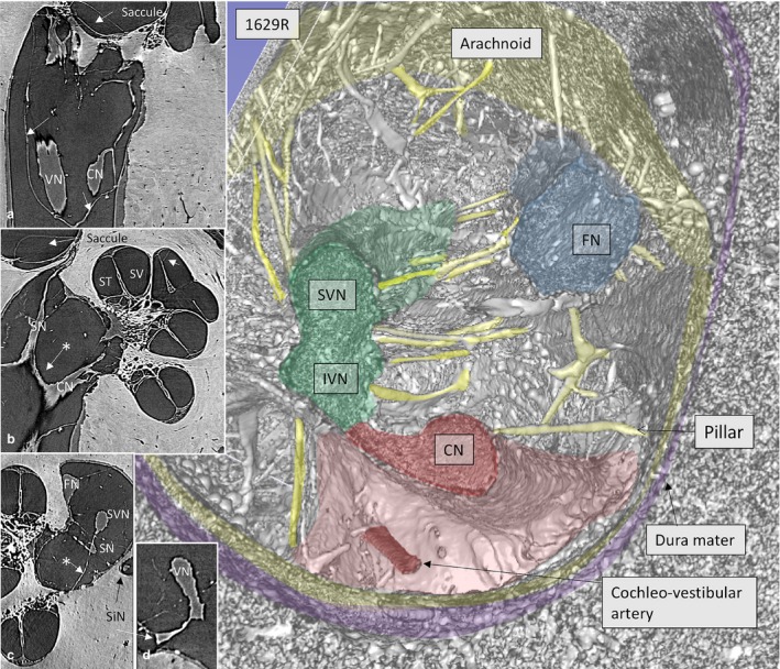
Figure 4.

SR‐PCI and 3D reconstruction show the nerve complexes in a left IAC surrounded by a continuous arachnoid sheet (inset a). The arachnoid is firmly attached to the dura wall (stained purple) except superiorly, where there is a subdural space. Inset b shows a section with connection between the CN and SN (*) as well as arachnoid attachments to the IAC walls. In inset c, the arachnoid connections between the SVN and SN and fundus can be seen (*). The cochleo‐vestibular artery (CVA) enters among the high‐frequency nerve fibers and reaches the base of the cochlea. ST, scala tympani; SV, scala vestibule.
