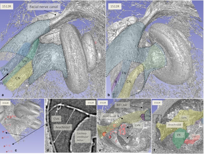
Figure 8.

(a,b) SR‐PCI and 3D reconstructions of a left human cochlea [medial view; (b) slightly angled anti‐clockwise]. The medial part of the IAC (blue) and cranial nerves are seen. (c–e) The cochleo‐vestibular artery (CVA) could be traced from the arachnoid in the IAC to the cochlea by fiducial markers (c,e). Arachnoid tags the IAC wall inferiorly and follows the entrance of the singular nerve (frame in f). CVN, cochleo‐vestibular nerve; FN, facial nerve; IN, intermediate nerve; SVN, superior vestibular nerve; VN, vestibular nerve. Small arrow; TC.
