Figure 1.

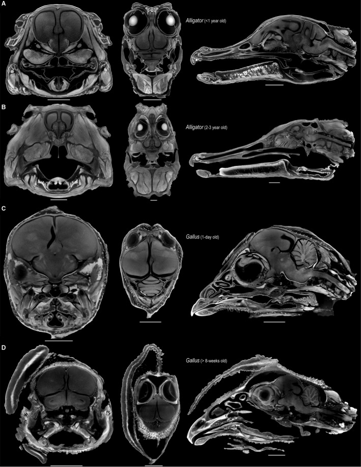
Selected transverse (left), frontal (middle), and sagittal (right) μCT slices through the heads of perinatal Alligator (A), 2‐ to 3‐year‐old Alligator (B), 1‐day‐old Gallus (C), > 8‐week‐old Gallus (D), illustrating the minimal shrinkage artifact from staining neural tissue with high concentrations of Lugol's iodine. Scale: 5 mm (A,C), 10 mm (B,D).
