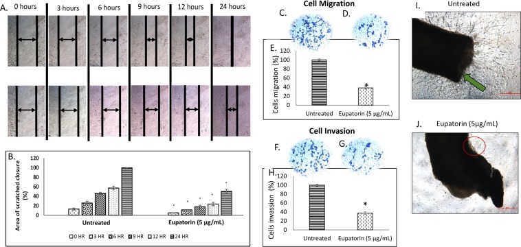
Figure 3.
(A) Representative image and (B) quantitative closure (%) for scratched assay. The confluent MDA-MB-231 cells were incubated with 5 μg/mL for the indicated times. The widths of injury lines made in cells were then examined at 0, 3, 6, 9, 12 and 24 hours. Images were taken using microscope (Nikon, Japan) at indicated periods. Magnification: x10; scale bar: 50 μm. Eupatorin inhibits serum-induced MDA-MB-231 cell (C–E) migration and (F,G) invasion in Boyden chamber assay after 24 hours. The pictures (C,D,F,G) show a section of the visual field of one representative experiment (x10 magnification). Scale bar: 50 μm. (C) migration of untreated MDA-MB-231 cells through membrane in the Boyden chamber assay, (D) migration of treated MDA-MB-231 cells, (E) percentage of migrated MDA-MB-231 cells, (F) invasion of untreated MDA-MB-231 cells, (G) invasion of treated MDA-MB-231 cells and (H) percentage of invaded MDA-MB-231 cells through Matrigel-coated membrane in the Boyden chamber assay. Statistical analysis was performed using unpaired t-test. (I) Untreated mouse aorta ring (J) eupatorin treated mouse aorta ring. Eupatorin inhibited the formation of new blood vessels in mouse aorta after 10 days incubation. Mouse aorta ring was viewed using Nikon microscope at x40 magnification. Scale bar: 100 μm. Green arrow indicated the formation of massive new blood vessels on aortic ring. Data are presented as mean values ± SD of n = 10 independent experiments. (*statistical significance (p < 0.05) against the untreated group).

