Figure 1.
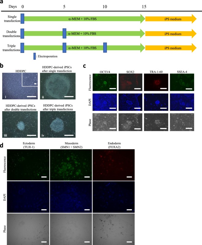
Generation of HDDPC-derived iPSCs. (a) Time-line for isolation of iPSCs from HDDPCs via single, double, or triple transfections. (b) Morphology of the HDDPCs (a) and HDDPC-derived iPSC colonies (b–d). iPSC colonies obtained after single (i), double (ii), or triple transfections (iii) from the HDDPC lines P02, P03, and P04, respectively. The magnified image is shown in the upper right portion of (i). Bar = 500 μm. (c) Immunocytochemistry of HDDPC (P01)-derived iPSC colonies using antibodies for OCT3/4, SOX2, TRA-1–60, and SSEA-4. Cells were stained by DAPI to visualise the location of nuclei. Bar = 500 μm. (d) Immunocytochemistry of embryoid body-derived outgrowth using antibodies for TUJI-1 (ectodermal marker), SMN1 + SMN2 (mesodermal marker), and FOXA2 (endodermal marker). Cells were stained by DAPI to visualise the location of nuclei. Bar = 500 μm.

