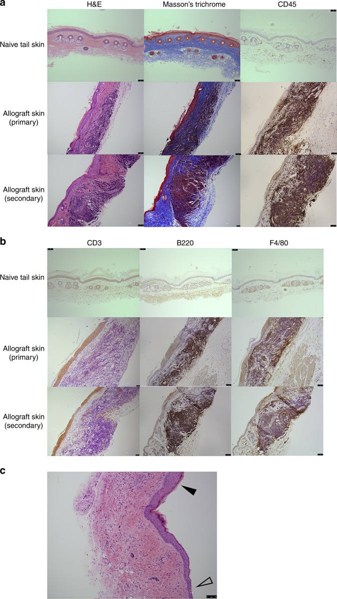
Fig. 3.
Hematopoietic cell infiltration of surviving skin allografts in CD117-ADC-conditioned recipient mice. a, b Untransplanted (naive) BALB/c tail skin (upper panels) and both primary (middle panels) and secondary (lower panels) skin allografts from the indicated mouse strains were sectioned and stained as indicated in the figure with H&E, Massonʼs trichrome, and CD45 (a) or CD3, B220, and F4/80 (b). Data are representative of 11 mice from two independent experiments. c Immune cell infiltration of surviving skin allografts from CD117-ADC conditioned mice does not extend into adjacent recipient skin. Cell infiltration at the edge of skin allografts (filled arrow head) and the surrounding recipient skin (open arrow head) is shown. Data are from H&E sections of skin and are representative of 11 mice from two independent experiments. Scale bars = 75 μm

