Fig. 1.
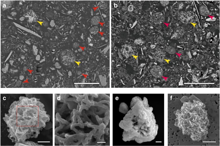
Representative electron micrographs of microparticles in sediment samples. a, b Cross-sectional scanning electron microscopy (SEM) images of resin-embedded oxic pelagic clay. Arrows indicate Mn-microparticles (yellow) and clay-microparticles (red) (samples U1365C-1H-2 0/20 and U1367D-1H-2 20/40, respectively). Scale bars, 10 μm. c SEM image of a Mn-microparticle in a density-separated sample (sample U1365C-1H-2 0/20). Scale bar, 5 μm. d Enlargement of the area within the red square in (c) showing the tangled fibrous strands in a microparticle. Scale bar, 500 nm. e, f SEM images of Mn-microparticles in density-separated samples (samples U1365C-9H-3 35/55 and U1366F-1H-2 40/60, respectively). Scale bars, 5 μm

