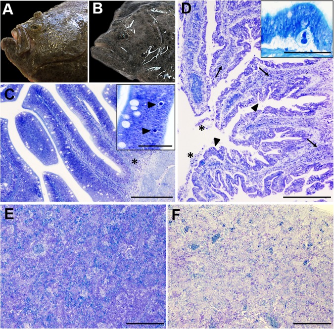
FIGURE 1.
Gross (A,B) and microscopic (C–F) pathology of turbot enteromyxosis. (A,B) Comparative photographs of a healthy turbot (A) and a specimen infected by Enteromyxum scophthalmi (B). Note the prominence of the head bony ridges in the diseased turbot associated to muscle atrophy, which constitute the characteristic external lesion of enteromyxosis named as “sunken head.” (C) Photomicrograph of turbot pyloric caeca at early stages of the parasitization. The intestinal architecture is maintained, subtle alterations such as slight mononuclear infiltrates (asterisk) and the presence of few early developmental stages of E. scophthalmi (arrowheads, inset) are observed. (D) Photomicrograph of turbot pyloric caeca at advanced stages of the parasitization. There is moderate to severe inflammatory infiltration of the lamina propria-submucosa (arrows) and presence of desquamated cells in the lumen (asterisks). The intestinal epithelium shows a typical “scalloped” shape (arrowheads) and harbors an elevated number of parasitic structures (see the inset, presenting a high magnification of a developmental stage 3 of E. scophthalmi). (E,F) Comparative photographs of the spleen in a control (E) and a severely infected (F) turbot, with an evident cell depletion in the latter. (C–E) Toluidine blue stain, bars = 200 μm (insets bars = 50 μm).

