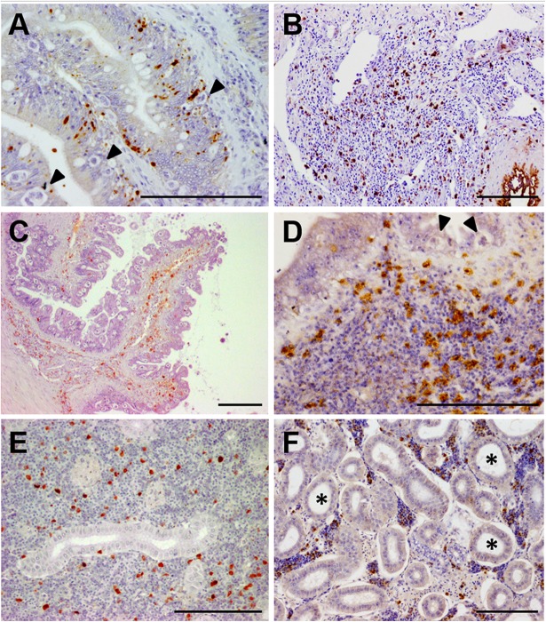
FIGURE 2.
Photomicrographs of illustrative immunohistochemical findings in E. scophthalmi-infected turbot. Brown color indicates positive staining, bars = 100 μm. (A) Immunostaining of active caspase-3, an indicator of apoptotic cell death, in the intestinal lining epithelium of turbot, where several parasitic structures are visible (arrowheads). (B) Inflammatory infiltrate in the lamina propria-submucosa of a diseased turbot, showing an abundant presence of cells immunoreactive to inducible nitric oxide synthase (iNOS). (C) Intestinal folds of a heavily parasitized turbot, where numerous IgM-positive (IgM+) cells can be observed, mainly in the lamina propria-submucosa, as well as an elevated parasitic load in the epithelium. (D) Immunoreactivity to tumor necrosis factor-alpha (TNFα) of several cells constituting the inflammatory infiltrate in the gut of an E. scophthalmi-infected turbot. Two parasites (arrowheads) are recognizable in the lining epithelium. (E) Turbot kidney showing scattered IgM+ in the lymphohematopoietic interstitial tissue of the organ. (F) Kidney of turbot with advanced enteromyxosis showing immunostaining to TNFα of some cells of the intertubular parenchyma, which suffered a remarkable cell depletion associated to dilatation of renal tubules (asterisks).

