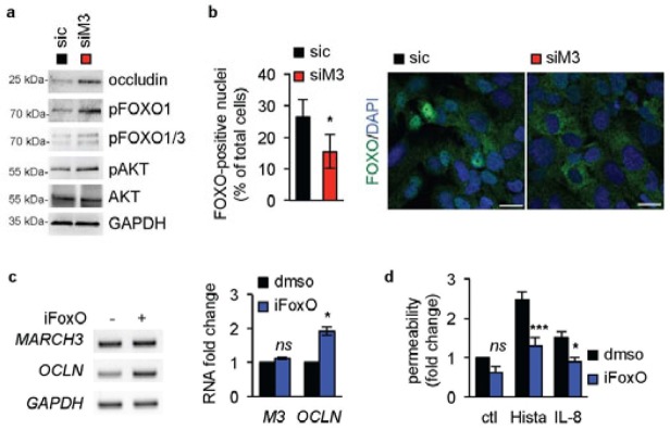
Figure 4. MARCH3 impact on OCLN gene expression relies on FoxO.
(A–B) Human brain endothelial cells were transfected with siM3 or sic duplexes, processed for western-blots three days later, as indicated. (B) Alternatively, cells were stained for FoxO1 (green) and analyzed by confocal microscopy. Nuclei were counterstained with DAPI (blue). Graph showed the percentage of FoxO1-positive nuclei. Scale bars: 10 μm. n>200 cells, T-test: *, p<0.05. (C) Cells were exposed to the FoxO1 inhibitor (AS1708727, iFoxo, 100 nM, three days) or vehicle (dmso) and processed for RT-PCR. (D) Permeability assays were performed in non-stimulated (ctl), histamine- (Hista) or IL-8-challenged HUVECs pre-treated with dmso and iFoxo. All panels are representative of at least three independents experiments. ANOVA test *, p<0.05; ***, p<0.001.

