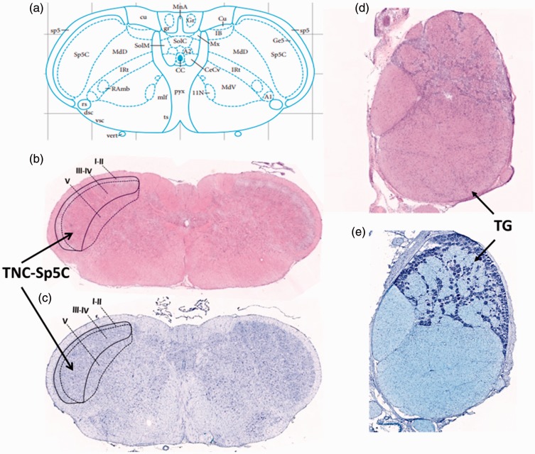
Figure 4.
Coordinates and structures of the TG and the TNC. (a) The stereotaxic coordinates of Sp5C in rat brain (adapted from Paxinos and Watson’s work). The H&E (b) and Nissl (c) staining of the TNC-Sp5C. The present study analyzed expression patterns of CGRP, PACAP, and its receptors in lamina III to V of the Sp5C. The H&E (d) and Nissl (e) staining of the TG.
TG: trigeminal ganglion; TNC: trigeminal nucleus caudalis; H&E: Hematoxylin and eosin; Sp5C :spinal trigeminal nucleus caudalis.

