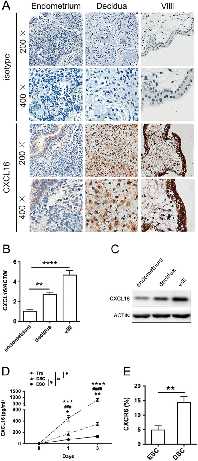
Figure 1.

Expression of CXCL16/CXCR6 at the maternal–foetal interface during decidualization. (A) Representative micrographs for CXCL16 immunostaining in normal endometrium from a control group (ii), decidual tissues (iii), and villus (iv) from normal pregnancies. Negative antibody isotype staining of the normal tissue was represented as control (i). Magnification, ×200, ×400. (B and C) The CXCL16 transcript and translation levels in endometrium, decidual tissues and villus were quantified by real-time PCR and immunoblotting and were then normalised to actin expression. (D) To identify the secretory CXCL16 level of ESCs, DSCs and trophoblast cells, the culture supernatants (1 and 3 days) were collected for CXCL16 measurements by ELISA. (E) The expression of CXCR6 in DSCs and trophoblast cells was detected in 3 days by flow cytometry. Decidua, decidua from normal pregnancy; DSC, decidual stromal cell from normal pregnancy; endometrium, normal endometrium; ESC, endometrial stromal cell; Tro, trophoblast cells from normal pregnancy; villi, villus tissues from normal pregnancy. Representative data were shown as well as the mean ± s.d. (n = 12). Scale bar = 50 mm. **P < 0.01, ****P < 0.0001. In (C), DSC vs ESC, *P < 0.05, **P < 0.01; Tro vs ESC, ###P < 0.001, ####P < 0.0001; Tro vs DSC, ★★★P < 0.001, ★★★★P < 0.0001.

 This work is licensed under a