Figure 5.
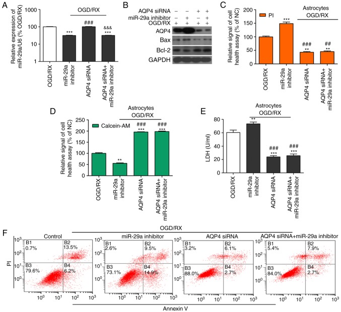
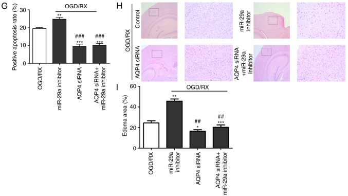
AQP4 mediates the protective effects of miR-29a in ischemia-induced astrocyte cell injury. (A) qRT-PCR determination of the expression of miR-29a in OGD/RX-induced astrocytes or NC. ***P<0.001 vs. NC; ###P<0.001 vs. miR-29 inhibitor, &&&P<0.001 vs. AQP4 siRNA. (n=3 for each experiment). (B) Western blot analysis of the expression of miR-29a in astrocytes treated with OGD for 6 h following different groups. All experiments were repeated in triplicate. Bands have been cropped from different parts of different gels, and respective western blot images have been cropped for Figure implementation. (C and D) Cell health assay of OGD-treated primary cultured astrocytes co-transfected with or without AQP4 siRNA and miR-29a inhibitor following PI/calcein staining. **P<0.01, ***P<0.001 vs. OGD/RX; ##P<0.01, ###P<0.001 vs. OGD/RX+miR-29a inhibitor (n=3 for each experiment). (E) Expression of LDH assessed by ELISA. **P<0.01, ***P<0.001 vs. OGD/RX; ###P<0.001 vs. OGD/RX+miR-29a inhibitor (n=3). (F and G) Apoptotic cells among OGD-induced astrocytes co-transfected with or without AQP4 siRNA and miR-29a inhibitor estimated by flow cytometry (n=3 for each experiment). **P<0.01, ***P<0.001 vs. OGD/RX; ###P<0.001 vs. OGD/RX+miR-29a inhibitor. (H) H&E detection of cell damage. (I) Bar graph of calculated brain oedema volume. *P<0.05, **P<0.01, ***P<0.001 vs. OGD/RX; ##P<0.01 vs. OGD/RX+miR-29a inhibitor. AQP4, aquaporin 4; OGD/RX, oxygen glucose deprivation-reoxygenation; NC, negative control; PI, propidium iodide.


