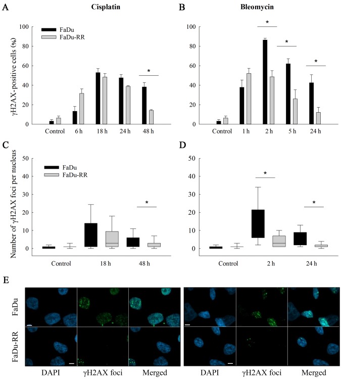
Figure 5.
DNA double-strand break repair after exposure to cisplatin or bleomycin. Bar charts represent percentage of γH2AX-positive radiosensitive (FaDu) and radioresistant (FaDu-RR) cells at different time points after exposure to cisplatin (A) or bleomycin (B). In the box plots, the number of γH2AX foci/nucleus at 2 time-points [18 and 48 h for cisplatin (C) or 2 and 24 h for bleomycin (D)] are shown. At each time-point at least 200 nuclei were analyzed. *P<0.05 between FaDu and FaDu-RR cell line. Images of γH2AX foci 24 h after exposure to chemotherapeutics were captured with a confocal microscope (Zeiss LSM 800; Carl Zeiss Microscopy GmbH, Jena, Germany), where nuclei stained with DAPI (blue) and γH2AX foci (green) are shown (E). Scale bar (white) represents 5 µm.

