. 2018 Nov 3;32(6):1387–1401. doi: 10.21873/invivo.11391

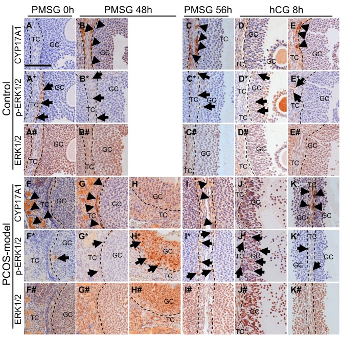
Figure 10. The localization of CYP17A1, phospho-ERK1/2 and ERK1/2 by immunohistochemistry in the ovary of 3-month-old control mice at diestrus(A, A* and A#) and PCOS-model mice (F, F* and F#). Control mice at 48 h after PMSG treatment (B, B* and B#), PCOS-model mice at 48 h afterPMSG treatment (G, G*, G#, H, H* and H#), control mice at 56 h after PMSG treatment (C, C* and C#) and PCOS-model mice at 56 h after PMSGtreatment (I, I* and I#) and control mice at 8 h after hCG treatment (D, D*, D#, E, E* and E#) and PCOS-model mice at 8 h after hCG treatment (J,J*, J#, K, K* and K#). Bar=100 μm. Arrowheads, CYP17A1 positive cells; arrows, phosho-ERK1/2 positive cells. n=4-6 in each group .

Figure 10