. 2018 Nov 3;32(6):1387–1401. doi: 10.21873/invivo.11391

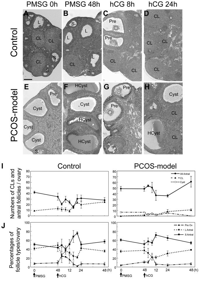
Figure 6. The histology of ovaries from 3-month-old control mice at diestrus (A) and PCOS-model mice (E). Bar=200 μm. Effects of PMSG and hCGinjections on the ovaries of 3-month-old control and PCOS-model mice. HE staining of 3-month-old control and PCOS model mouse ovaries at 0 (A,E), and 48 h after PMSG (B, F), 8 (C, G) and 24 h (D, H) after hCG. S, small antral follicle; L: large antral follicle; Pre: preovulatory follicle, CL:corpus luteum, HCyst: hemorrhagic cyst, Cyst, cyst. The total numbers of antral follicles, corpora lutea (CLs) and cysts in ovaries from control andPCOS-model mice were counted at 0 and 48 h after PMSG injection, and 4, 8, 12, 24 and 48 h after hCG injection, respectively (I and J). Folliclestage index, preovulatory (>390 μm), large antral (250-390 μm) and small antral follicles (140-250 μm), respectively. n=5 in each group.

Figure 6