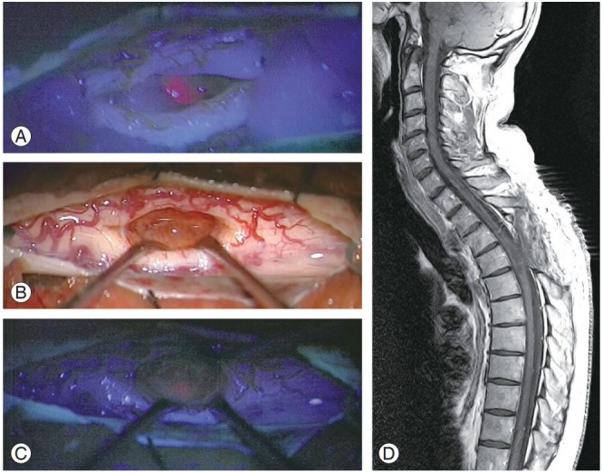
Fig. 1.

Case 4. (A) Intraoperative image with blue light showing the intense fluorescence emitted by the lesion. (B) Intraoperative image with white light in which the tumor bed is seen. (C) Intraoperative image with blue light in which no fluorescence emission is seen in the tumor bed. (D) Sagittal dorsal postoperative magnetic resonance imaging shows complete resection.
