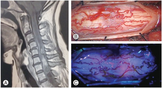
Fig. 2.

Case 7. (A) Sagittal cervicodorsal magnetic resonance imaging with gadolinium shows the lesion. (B) Intraoperative image with white light shows the spinal cord with numerous pathologic vessels on its surface. (C) Intraoperative image with blue light shows fluorescence emission before myelotomy.
