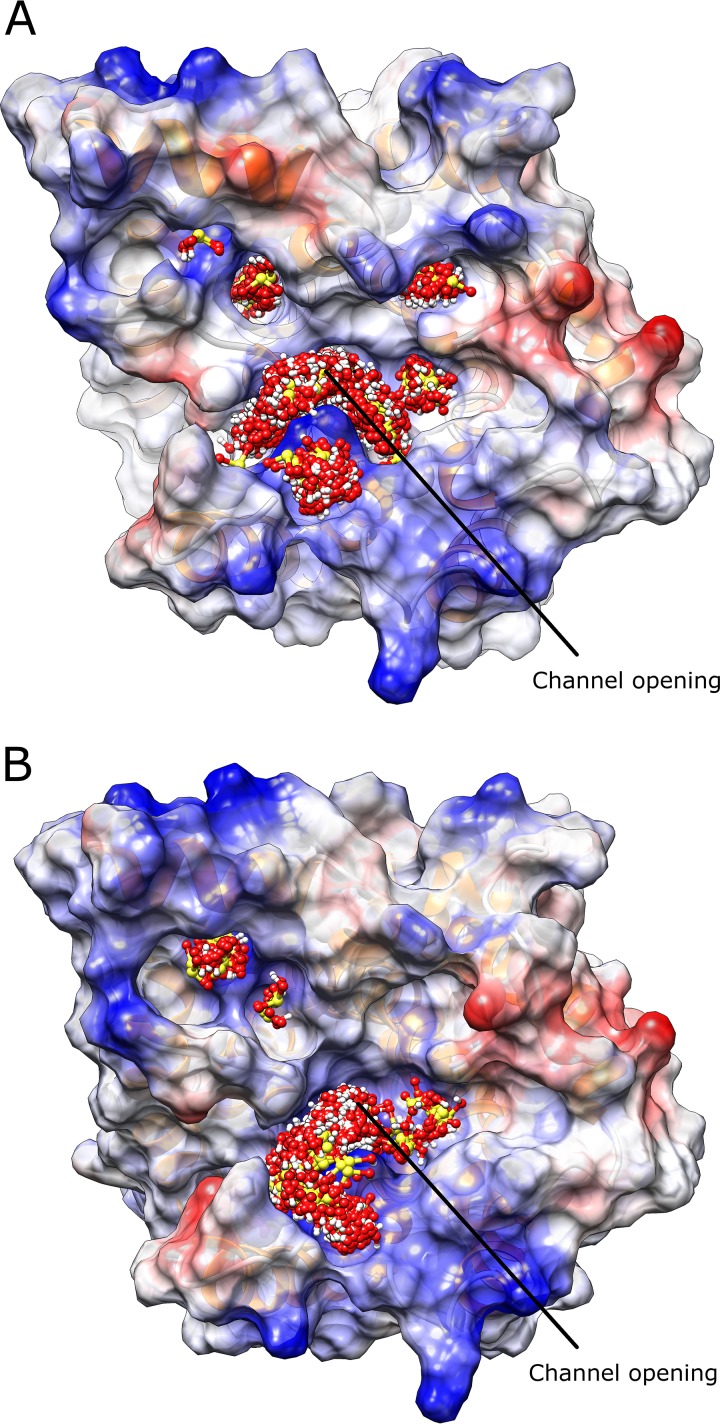
FIG 7.
HSO3− docking simulations for protein structures of AWRI1499_SSU1_hap2 (A) and AWRI1613_SSU1_hap2 (B). Protein structures are shown from the cytoplasm with protein molecular surface colored by Coulombic electrostatic potential (blue, positive charge; red, negative charge). HSO3− is colored by atom (red, oxygen; white, hydrogen; yellow, sulfur).

