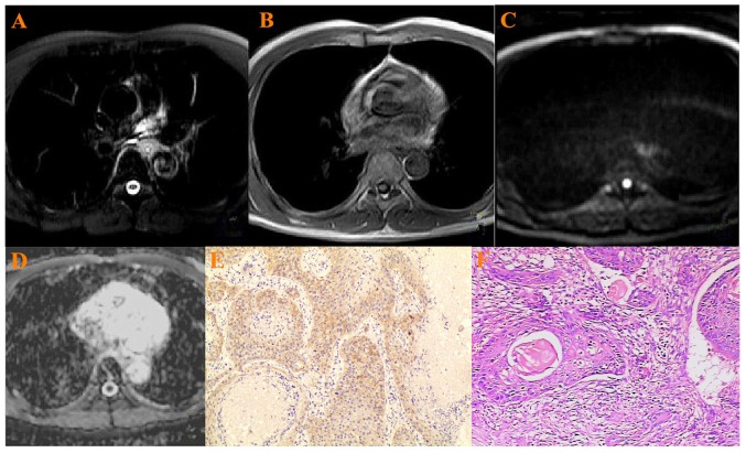
Figure 1.
A 75-year-old patient with ESCC in the mid-thoracic portion. (A) ESCC was hyperintense on T2-weighted image, (B) hypointense on T1-weighted image, (C) hyperintense on the DW-MRI (b=800 s/mm2) (D) hypointense on ADC map. ADC value:1.62±0.35×10⁻³ mm2/s. (E) Low VEGF staining in cytoplasm of ESCC: Weak positive (+) (×200 magnification), (F) Histopathology: Well differentiated squamous cell carcinoma (×200 magnification). ESCC, esophageal squamous cell carcinoma; DW-MRI, diffusion weighted-magnetic resonance imaging; ADC, apparent diffusion coefficient; VEGF, vascular endothelial growth factor.

