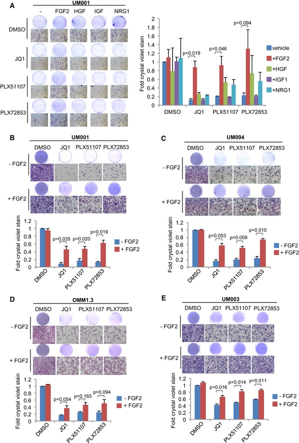
(A) UM001 cells were treated with 1 μM JQ1, 1 μM
PLX51107 or 100 nM
PLX72853 in combination with 50 ng/ml FGF2, 50 ng/ml HGF, 50 ng/ml IGF1 or 50 ng/ml NRG1 for 8 days. Changes in cell viability were determined by crystal violet staining. FGF2 rescued (B) UM001, (C) UM004, (D) OMM1.3 and (E) UM003 from the growth inhibitory effects of BET inhibitors. UM004 cells were treated with 2 μM JQ1, 2 μM
PLX51107 or 200 nM
PLX72853 in combination with 50 ng/ml FGF2 for 8 days. Other cells were treated with 1 μM JQ1, 1 μM
PLX51107 or 100 nM
PLX72853 in combination with 50 ng/ml FGF2 for 8 days. Cell growth was determined by crystal violet staining. Data presented are fold change in crystal violet stain compared to untreated control and are mean ± SEM from triplicate experiments (or
n = 3). The unpaired
t‐test was used for statistical significance. Representative crystal violet images are shown. Scale bar: 100 μm.

