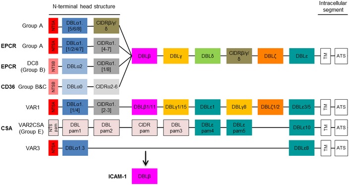
Figure 1. PfEMP1 domain structure.

A schematic presentation of PfEMP1 domain structure comprising a N‐terminal head structure, 2‐6 subsequent C‐terminal domains, a transmembrane domain (TM) and an intracellular acidic terminal segment (ATS) with known receptors indicated in bold. Receptor specificity is determined by combined DBL and CIDR domains with mutually exclusive binding to EPCR and CD36 by different CIDRα domains in the head structure. Part of group A PfEMP1 and a particular subset of group B/A chimeric PfEMP1 (DC8) bind to EPCR via CIDRα1 domains, whereas group B and C PfEMP1 bind CD36 via CIDRα2‐6 domains. The atypical group E VAR2CSA PfEMP1 binds placental chondroitin sulphate A (CSA) via DBLpam1 and DBLpam2 domains. The binding phenotype of VAR 1, VAR3 and group A CIDRβ/γ/δ domains is unknown, but they do not bind EPCR or CD36. DBLβ domains can be involved in ICAM‐1 binding and are from both groups A and B. Not much is known about the other DBL domains (γ/δ/ε/ζ), but the DBLε and DBLζ domains are implicated in IgM and α2‐macroglobulin binding.
