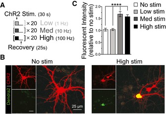
Figure 4. Increased neuronal activity increases non‐AUG‐dependent translation of DPRs in primary cortical neurons.

-
ANeuronal activity was induced with Chr2 stimulation by performing trains of LED light pulses at various frequencies: No stim = 0 Hz; Low = 1 Hz; Med = 10 Hz; High = 100 Hz. Trains included 30 s of light pulses followed by 25 s of recovery for desensitized ChR2; trains were performed 20 times.
-
BMicroscopy images show increased DPR fluorescent intensity levels from medium‐ and high‐frequency stimulation of ChR2 in primary cortical neurons transfected with the C9 DPR reporter.
-
CRelative fluorescent intensity quantification shows a significant increase in DPR levels with increases in neuronal activity through medium and high stimulation in primary cortical neurons. No stimulation and low stimulation frequencies did not increase levels of DPR. The C9 DPR reporter, monitoring the GP DPR (ORF2), was cotransfected with ChR2. Cortical neuron: n = 4 with m > 10 cells per n. (****P < 0.0001). All error bars represent the SEM, and all statistics were performed using an uncorrected Fisher's exact test.
