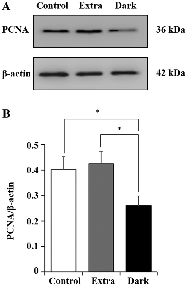
Figure 3.
PCNA expression in maple syrup-treated DLD-1 cells. (A) Western blot analysis indicated the expression levels of PCNA in DLD-1 cells treated in the control group (without maple syrup) and in the two maple syrup groups, the Extra and Dark groups. (B) Quantification of western blot analysis results. Data is presented as the mean ± standard error of the mean of three independent experiments in maple syrup-treated DLD-1 cells. *P<0.05. PCNA, proliferating cell nuclear antigen; Extra, extra light maple syrup; Dark, dark-colored maple syrup.

