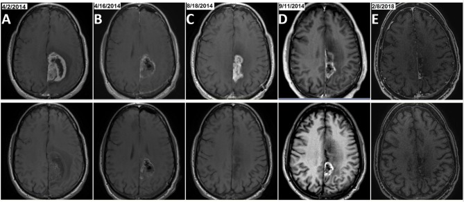
Figure 3.
Example of MRI results from patient number 3, a long-term survivor in the TBI+T group. The patient was treated with initial craniotomy and concurrent radiation and temozolomide followed with maintenance TMZ. He was treated with TBI (22 treatments with BEV and IRI from March 2015 to January 2016) after his first GBM progression (rGBM) was resected. The patient finished a cumulative 30 cycles of TMZ when the TBI regimen was completed and he continued on TTFields treatment alone for 39 months as of March, 31, 2018. Two craniotomy surgeries were performed in April and September of 2014, respectively. Pre- and post-craniotomy MRIs, T1 with contrast for newly diagnosed GBM [(A), 4/2/2014 and post-surgery at (B), 4/16/2014] and rGBM [(C), 8/18/2014 and post-surgery at (D), 9/11/2014] as well as the most recent brain MRI immediately prior to the data cut-off date [(E), 2/8/2018] are shown in upper row, with brain MRI T1 without contrast shown in the corresponding lower row.

