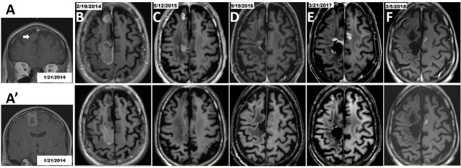
Figure 4.
Therapy with TBI in combination with Gamma Knife stereotactic radiosurgery after the second GBM progression in patient number 5. Initial craniotomy was on 1/25/2014, but axial section images were not available in the pre-operative MRI. (A) Coronal view of anterior right frontal lesion (arrow). (A') Coronal view of posterior right frontal lesion. (B) Post-operative MRI. Tumor in the posterior right frontal lobe was removed while the mass at the right anterior frontal lobe was untouched. (C) MRI on 5/12/2015 showed that tumor in the right anterior frontal lobe progressed as a non-targeted lesion while on EF-14 clinical trial, and this lesion was treated with Gamma Knife on May 20, 2015. The patient had been treated with TBI therapy for 1 year and then it stopped as scheduled. (D) MRI on 9/19/2016 suggested tumor progression around the surgical cavity and he had a 2nd craniotomy on 9/25/2016. But pathology demonstrated no active tumor. This was not considered as progression of disease. (E) The 2nd disease progression was diagnosed based on MRI (3/21/2017) because of an appearance of a new lesion in the left hemisphere which was treated with Gamma Knife on 4/5/2017. The TBI regimen was restarted. (F) The progressive disease was well-controlled based on the latest MRI on 3/5/2018; lesions were treated with Gamma Knife followed by TBI regimen plus TTFields. Brain MRI T1 with contrast shown in upper row and brain MRI T1 without contrast shown in the corresponding lower row.

