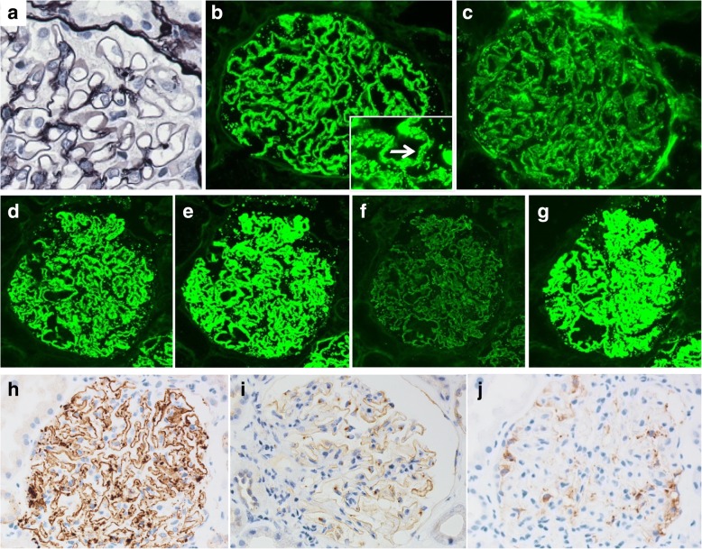
Fig. 1.
Microscopic findings of the glomeruli in the kidney. Periodic acid-methenamine silver staining shows no obvious abnormality (a). Immunofluorescence shows granular staining for IgG (b, arrow), C3 (c), IgG1 (d), IgG2 (e), IgG4 (g) along the capillary wall with weak staining for IgG3 (f). Immunohistochemical staining shows strong staining for THSD7A along the capillary wall in the case (h), in normal control (i), with negative staining for PLA2R in the case (j)

