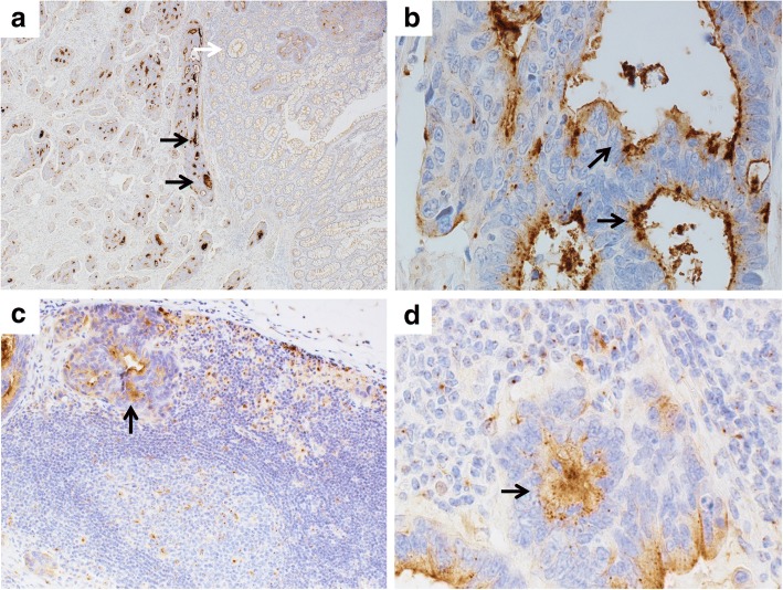
Fig. 3.
THSD7A staining in the rectal cancer, and metastatic lymph node. Strong staining for THSD7A is shown in tumor tissue (a, black arrows), with no staining in non-tumor tissue (a, white arrow). THSD7A shows luminal pattern staining in tumor tissue (b, arrows). THSD7A staining is shown in metastatic cell in lymph node (c, d, arrows). Original magnification a: × 40, b: × 600, c: × 200, d: × 600

