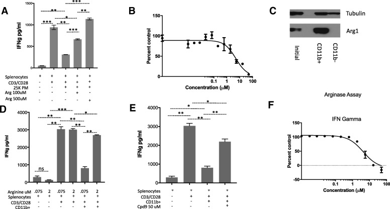
Fig. 3.
M2 macrophages and Cd11b + cells express Arginase 1 and suppress T cell function. a Cpd9 50 uM prevented M2 peritoneal macrophages suppression of IFNg secretion by splenocytes. Splenocytes were activated with CD3/28 in the presence of 25,000 M2 peritoneal macrophages supplemented with arginine. IFNg was determined by ELISA in the culture supernatants after 24 h. Splenocytes 50K, PM 25K. b IC50 of Cpd9 in 15K M2 peritoneal macrophages. c Western blot analysis of Arginase 1 expression in CD11b + cells. CD11b + cells were sorted from ID8 tumor ascites fluid, and Arginase 1 expression was characterized by western blot using Arginase 1 and tubulin antibodies. d Arginine 2 mM prevented CD11b + cells suppression of IFNg secretion by splenocytes. Splenocytes were activated with CD3/28 in the presence of 0.075 mM or 2 mM arginine as indicated. Sorted CD11b + cells were added to the indicated wells. After 24 hs IFNg was determined by ELISA in the culture supernatants. Splenocytes 50K; CD11b: 150K e. Cpd9 50 uM prevented CD11b + MDSC cells suppression of IFNg secretion by splenocytes. Splenocytes were activated with CD3/28 in the presence of arginine 75 uM, sorted CD11b + MDSC and Cpd9 50 uM were added to the indicated wells. IFNg was determined by ELISA in the culture supernatants after 24 h. Splenocytes 50K; CD11b: 150K f IC50 of Cpd9 in CD11b MDSC suppression of splenocytes IFNg secretion. Splenocytes were activated with CD3/28 in the presence of arginine 75 uM arginine, sorted CD11b + MDSC and various concentrations of Cpd9. After 24 hs IFNg was determined by ELISA in the culture supernatants and the % of control (no Cpd9) was calculated. Splenocytes 100K, Arginase 0.35 μg/ml, and Arginine 75 uM. *p < =0.05, **p < =0.01, and ***p < =0.001. Graphs show representative results from experiments performed at least three times

