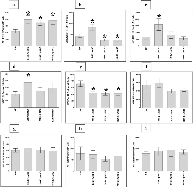
Fig. 7.
Flow cytometric analysis of indicated inflammatory molecules by NK cells harvested from NK/pMSC cytolytic experiments. As compared to untreated NK cells, pMSCs significantly increased NK cell expression of IL-18 Rβ at all indicated NK to pMSC ratios, *P < 0.05 (a); significantly increased NK cell expression of IL-12 Rβ1 (b), IFN-ɣ R1 (c), and TLR3 (d) at 25:1 NK to pMSC ratio, *P < 0.05; significantly decreased NK cell expression of IFN-ɣ R2 at all indicated NK to pMSC ratios, *P < 0.05 (e); and significantly decreased NK cell expression of IL-12 Rβ1 at 50:1 and 100:1 NK to pMSC ratios, *P < 0.05 (a). pMSC had no significant effect on NK cell expression of IFN-ɣ R1 (c) and TLR3 (d) at 50:1 and 100:1 NK to pMSC ratios, P > 0.05, and on IL-18 Rα (f), TNF-α (g), TLR7 (h), and TLR9 (i) at all indicated NK to pMSC ratios, P > 0.05. Experiments were carried out in triplicate and repeated for ten times using ten independent NK/pMSC cytolytic experiments where in each experiment NK cells, and pMSCs were prepared from the peripheral blood of ten different healthy subjects and ten different normal human term placentae, respectively. Bars represent standard errors

