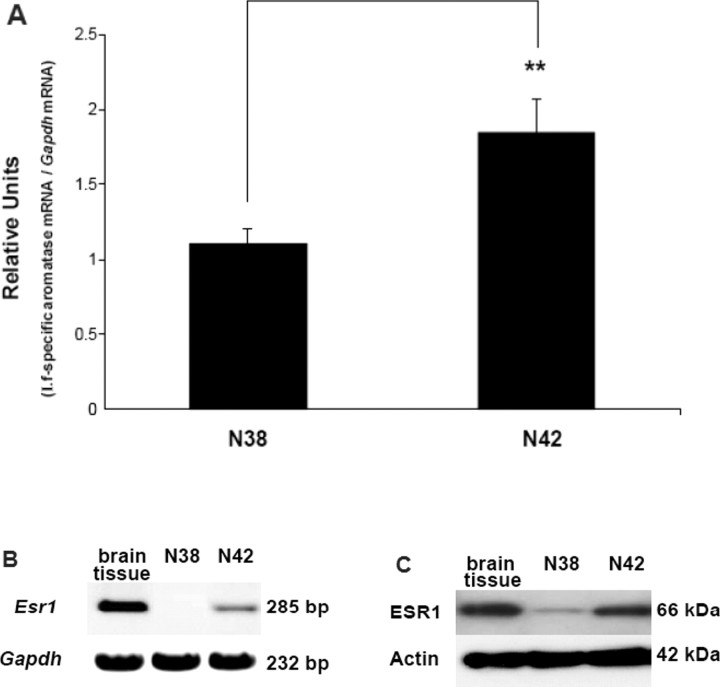
Fig. 1.
N42 and N38 hypothalamic cells use promoter I.f to regulate aromatase mRNA expression. Esr1 mRNA and ESR1 protein expression levels in N42 hypothalamic cells are higher than N38 hypothalamic cells. A) Real-time RT-PCR was performed employing a probe complementary to the exon I.f-exon II junction to measure promoter I.f-driven aromatase mRNA expression. Relative units are shown as the ratio of aromatase mRNA:Gapdh mRNA; (presented as l.f-specific aromatase mRNA/Gapdh mRNA). Results are expressed as the mean ± SEM (n = 3); **P < 0.01, paired t-test. Conventional RT-PCR (B) and IB (C) were performed to measure ESR1 expression in brain tissue (positive control) and N42 or N38 hypothalamic cells. Gapdh and actin were used as loading controls. The figures represent one of three independently performed experiments.

