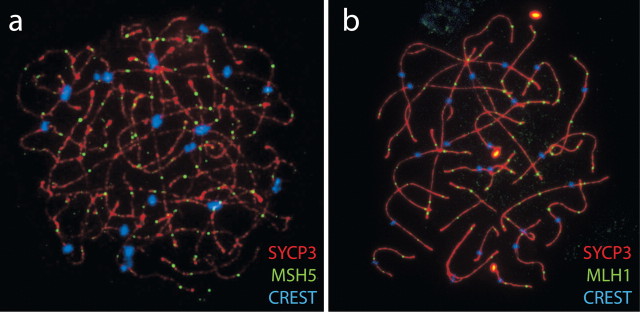
Fig. 2.
Examples of human oocyte chromosome spreads. Chromosome spreads were prepared from 21-wk-old fetal ovaries and immunostained against MutS protein homolog 5 (MSH5; a) and MutL homolog 1, colon cancer, nonpolyposis type 2 (MLH1; b), both in green, to reveal sites of recombination processing. In both images, the synaptonemal complex is highlighted in red by the use of anti-SYCP3 antibodies, whereas the centromere is stained blue with CREST (calcinosis, Raynaud phenomenon, esophageal dysfunction, sclerodactyly, and telangiectasia) human autoimmune serum. Methods are similar to those described previously [6]. In the absence of the HFOnet, such immunofluorescent techniques are one of only a few techniques available for studying regulators of meiotic function in fetal oocytes. This technique requires prior knowledge of the proteins of interest but can be used to validate and confirm information obtained from the HFOnet. Original magnification ×240.

