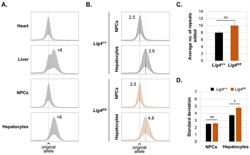
Fig. 2. Expansions in liver are confined to hepatocytes.

A. and B. Repeat PCR profiles from 3-month old males with 170 repeats. The black dotted line in each panel indicates the size of the original inherited allele, while the gray dotted line indicates the average size of the expanded alleles. A. Representative profiles of heart, liver, NPCs and hepatocytes showing the number of repeats added relative to the original allele. B. Representative profiles of NPCs and hepatocytes isolated from Lig4+/+ and Lig4fl/fl mice livers showing the SD and the calculated normal distribution profile of each allele. C. and D. show the average of repeats added and SDs respectively for 3 Lig4+/+ and 4 Lig4fl/fl animals with ~170 repeats ± SE. * p< 0.005, ns: not significant.
