
Fig. 3. Hepatocyte ploidy does not affect repeat instability.
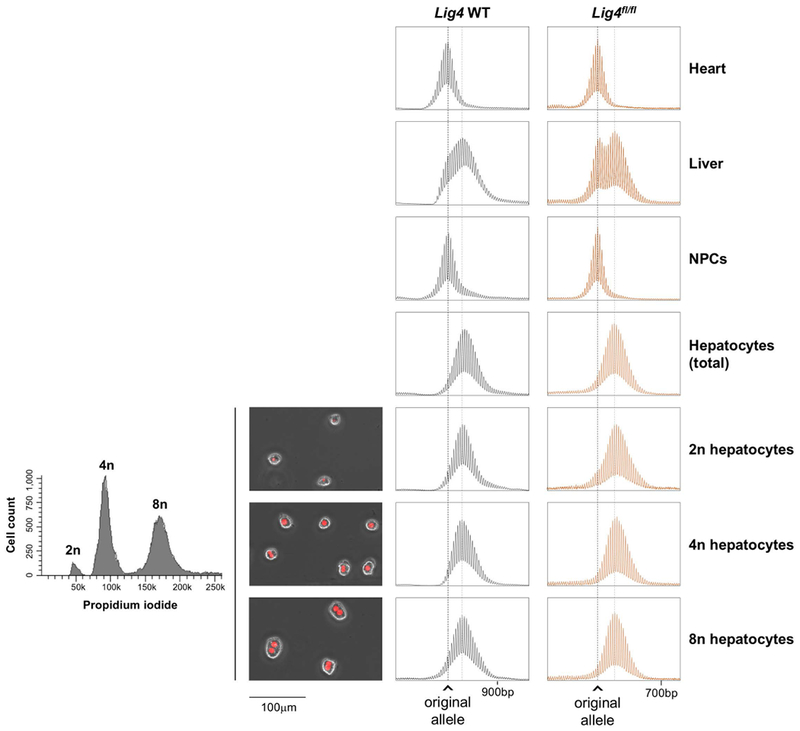
The chart on the left depicts the result of the cell sorting showing the proportion of the diploid and polyploid fractions as assessed by DNA content revealed by PI staining. The adjacent photomicrographs show typical examples of the indicated cell types, with propidium iodide (PI) in red. The right-hand side of the slide shows the Repeat PCR profiles of heart, liver, NPCs, total hepatocytes and diploid and polyploid hepatocyte fractions from a 2-months old Lig4 WT mouse with 260 repeats and a 3-month old Lig4fl/fl mouse with 170 repeats. The black dotted line in each panel indicates the size of the original inherited allele, while the gray dotted line indicates the average size of the expanded alleles.