FIGURE 2.

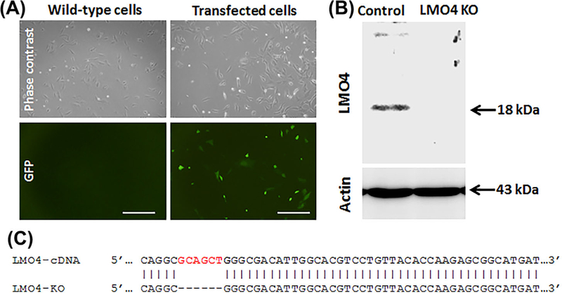
Knockout of LMO4 in UB/OC1 cells. A, Green fluorescent cells indicate the presence of GFP in UB/OC1 cells transfected with CRISPR/Cas9 knockout plasmid. GFP is not detected in the wild-type cells. The images are representative of three biological replicates. Scale bar represents 200 μm. B, Immunoblots indicate that LMO4 protein is not detected in the knockout cells. Actin is used for normalization. The images are representative of three biological replicates. C, Modified DNA sequence of target locus in the knockout cells indicates a 6 base pair deletion.
