Fig. 4.
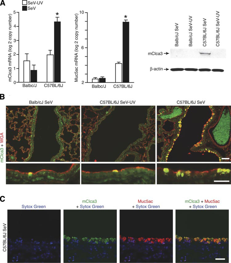
Detection of mClca3 gene expression in concert with mucous cell metaplasia in F0 parental mice. Balb/cJ and C57BL/6J mice were inoculated with SeV or SeV-UV and then analyzed as follows. A: lung RNA from 21 days after inoculation was subjected to real-time PCR for mRNA levels of mCcla3 and Muc5ac normalized to the Gapdh control. Values represent means ± SE for 4–6 mice. *Significant difference from SeV-UV. B: corresponding lung lysates from C57BL/6J mice were subjected to Western blot analysis with anti-human (h)CLCA1/ mClca3 Ab and control anti-β-actin mAb. C: corresponding lung sections from C57BL/6J mice were immunostained with anti-hClca1/mClca3 Ab and FITC-conjugated secondary Ab as well as anti-wheat germ agglutinin (WGA) Ab and CY3-conjugated secondary Ab. D: corresponding lung sections were immunostained with anti-hCLCA1/mClca3 Ab and Alexa 633-conjugated secondary Ab (green) as well as biotinylated anti-Muc5ac Ab and Alexa 555-streptavidin (red) and counterstained with Sytox green (blue). Bars = 20 μm.

