Image 1.
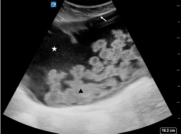
Point-of-care ultrasound performed with a curvilinear probe in the right lower quadrant shows a large, anechoic (black) collection of cerebrospinal fluid (white star) encapsulated by a fibrous layer (white arrow) and containing echogenic debris and hyperechoic (white) septations (black arrowhead).

