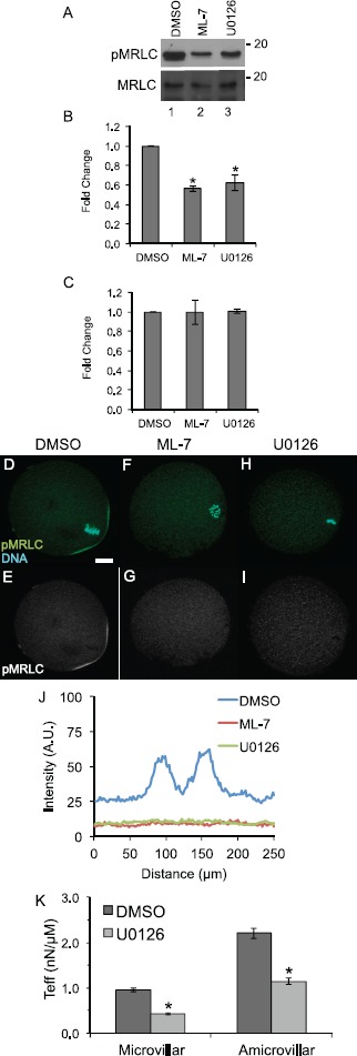
Fig. 1.

MAPK3/1 and MLCK inhibition reduces levels of active, phosphorylated myosin regulatory light chain, affects localization of phosphorylated myosin regulatory light chain, and decreases cortical tension in metaphase II eggs. A) Representative blots of egg lysates (90 cells per lane treated for 90 min with 0.5% DMSO, lane 1, 15 μM ML-7, lane 2, or 50 μM U0126, lane 3) probed with anti-pMRLC and anti-MRLC. B and C) Quantification of band intensities of anti-pMRLC levels (B) and anti-MRLC levels (C). Values were normalized to DMSO-treated eggs. Error bars represent standard errors of the mean. The anti-pMRLC blots were performed 13 times for DMSO-treated eggs, 11 times for ML-7-treated eggs, and eight times for U0126-treated eggs. The difference between levels of pMRLC in ML-7- and U0126-treated eggs is statistically significant as compared to DMSO-treated eggs (Kruskal-Wallis test with Bonferroni-Dunn post hoc testing, *P < 0.05). The MRLC blots were repeated four times for all the treatment groups. D–I) Immunofluorescence shows the localization of anti-pMRLC staining (green, with complementary black-and-white image of this channel in E, G, and I) and DNA (stained with DAPI, in blue) in metaphase II eggs exposed to 0.5% DMSO (D, E), treated with 15 μM ML-7 (F, G), or with 50 μM U0216 (H, I). Bar in D = 10 μm. J) Line-scan analysis around the circumference of the cortical region to assess relative intensities of the pMRLC cortical signal in DMSO-, ML-7-, and U0126-treated eggs. This graph shows pooled data from 23 DMSO-treated metaphase II eggs (blue line), 12 ML-7-treated eggs (red line; four metaphase II, six metaphase II drifted spindle, two anaphase II), and 18 U0126-treated eggs (green line; eight metaphase II, six metaphase II drifted spindle, and four anaphase II); see also Supplemental Figure S1 for data on individual eggs. In DMSO-treated eggs, the two peaks in these scans coincide with pMRLC at the two boundaries of the amicrovillar domain. The line scans of ML-7- and U0126- treated eggs do not show these peaks. K) Effective tension (Teff, in nN/μm) on the microvillar and amicrovillar domains of DMSO-treated eggs (dark gray) and U0126-treated eggs (light gray) as measured by micropipette aspiration. Error bars represent standard errors of the mean. This decrease in effective tension is statistically significant (Mann-Whitney U-test, *P < 0.05). Numbers of eggs analyzed: DMSO-treated microvillar domain, 36; U0126-treated microvillar domain, 79; DMSO-treated amicrovillar domain, 18; and U0126-treated amicrovillar domain, seven.
