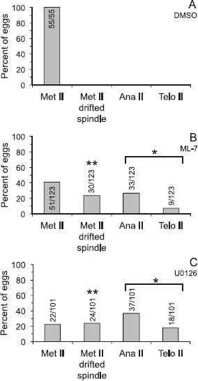
Fig. 3.

Effects of U0126 or ML-7 treatment in metaphase II eggs. Metaphase II eggs were treated with 0.5% DMSO (A), 15 μM ML-7 (B), or 50 μM U0126 (C) for 3 h, then fixed and stained with DAPI to label DNA, anti-β-tubulin to label the meiotic spindle, and the monoclonal antibody MPM-2 to label mitotic phosphoproteins (analysis shown in Fig. 5). Eggs were classified as metaphase II (Met II; normal metaphase II spindle morphology and cortical localization), Met II drifted spindle (i.e., the DNA was aligned along the metaphase II plate and the spindle was greater than 12 μm from the cortex; shown in Fig. 2), anaphase II, or telophase II (Ana II and Telo II, respectively, i.e., progressing out of metaphase II arrest). Numbers in or above bars indicates numbers of eggs analyzed. The extent of progression out of metaphase II arrest (combined numbers of eggs in anaphase II and telophase II states) in U0126- and ML-7-treated eggs is statistically significant as compared to DMSO-treated eggs (chi-square analysis, *P < 0.0001). The extent of drifted spindle incidences in U0126- and ML-7-treated eggs is statistically significant as compared to DMSO-treated eggs (chi-square analysis, **P < 0.0001).
