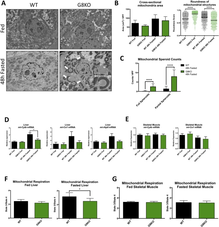
Figure 4.
Mitochondrial dysmorphology and reduced hepatic oxidative function in fasting GLUT8-deficient mice. (A) Electron micrographs of livers of WT and G8KO fed or fasted mice with identification of mitochondrial spheroids (inset, white arrows). (B) Quantification of mitochondrial cross-sectional area and roundness from electron micrographs. (C) Enumeration of mitochondrial spheroids in electron micrographs from fasted mice. Komogorov-Smirnov test was used to analyze distribution of roundness scores. (D) qRT-PCR analysis of hepatic mitochondrial DNA-encoded mRNA for ETC proteins (n = 6 mice per group). A significant two-way analysis of variance diet-genotype interaction was detected for mt-Cytb (P < 0.05). (E) qRT-PCR analysis of skeletal muscle mitochondrial DNA-encoded mRNA for ETC proteins (WT fed, n = 4; G8KO fed, n = 4; WT fasted, n = 8; G8KO fasted, n = 8). (F) Quantification of oxygen consumption in liver homogenates from fed or 48-hour–fasted WT and G8KO mice using the Oroboros Oxygraph 2K. Respiration is reported as the state 3/state 4 respiratory control ratio and subjected to paired t test analysis (n = 4 to 6 mice per group). (G) Quantification of oxygen consumption in permeabilized soleus muscle fibers from fed or 48-hour–fasted WT and G8KO mice (n = 4 to 6 mice per group). Data are presented as mean ± standard error of the mean. *P < 0.05; ****P < 0.0001.

