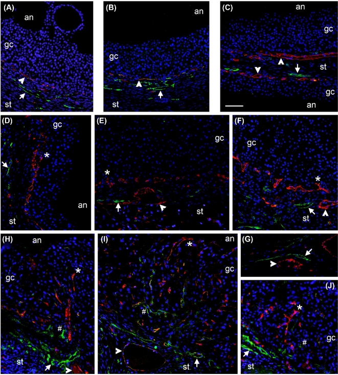
Figure 3.
Angiogenesis, but not lymphangiogenesis, precedes ovulation in primate follicles. Immunofluorescent detection of vascular endothelial cells (VWF, red), lymphatic endothelial cells (LYVE1, green), and cell nuclei (DAPI, blue) was performed in monkey ovarian tissues collected before hCG (A–C, 0 h hCG) or 36 h after hCG (D–F) but prior to ovulation. Image in panel C shows two adjacent follicles with intervening stroma (st). Ovaries were also collected during natural menstrual cycles 2 days after peak serum LH and visual observation of an ovulatory stigmata at the time of collection (H–J). In all panels, arrowheads indicate VWF+ cells in the perifollicular stroma, arrows indicate LYVE1+ cells in the perifollicular stroma, asterisks (*) indicate VWF+ cells among granulosa/granulosa-lutein cells, and hash signs (#) indicate LYVE1+ cells among granulosa/granulosa-lutein cells. (G) Detection of vascular endothelial cells (VWF, red, arrowhead) and lymphatic endothelial cells (LYVE1, green, arrow) in large vessels of the ovarian medulla served as a positive control. Ovarian stroma (st), granulosa/granulosa-lutein cells (gc), and follicle antrum (an) are indicated. Images are representative of n = 3–4 animals/group and are at the same magnification; bar in panel C = 50 μm.

