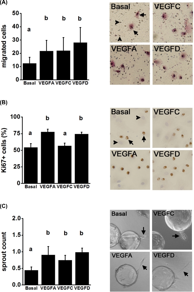
Figure 6.
VEGF family members promote angiogenic events in vitro. (A) Migration of mOMECs through a porous membrane in response to treatment for 24 h with basal medium alone or with the addition of VEGFA, VEGFC, or VEGFD. Representative images of migrated cells after each treatment. In basal image, arrows indicate migrated cells (pink); arrowheads indicate pores. (B) Proliferation of mOMECs as assessed by Ki67 immunodetection and expressed as percentage of total cells which were Ki67+. Representative images for each treatment are shown; examples of nuclei which are Ki67+ (brown, arrows) and Ki76– (blue, arrowheads) are indicated in basal image. (C) Sprout formation by mOMECs after culture with each treatment for 2 days. Representative images show beads with mOMECs forming sprouts (arrows) on day 2 in vitro for each treatment. For each graph, data were assessed by ANOVA and the Duncan test; groups with no common letters are different, P < 0.05; n = 4 mOMEC lines/group.

