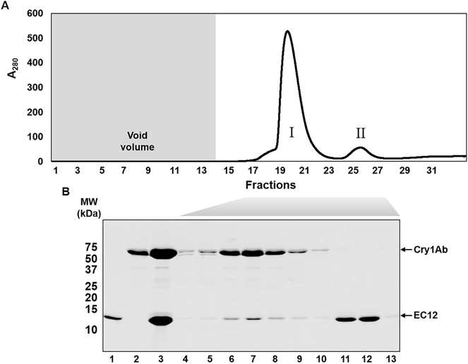
Figure 1:
Formation of the CrylAb toxin-EC12 complex in solution. CrylAb toxin was extracted from sporulated cells of B. thuringiensis subsp. berliner 1715. Purified toxin and EC12 of the receptor BT-R1 were mixed together and subjected to size exclusion chromatography (SEC) using a Superdex 75 10/300 GL column (see Materials and Methods). (A) Chromatogram for separation of the Cry1Ab-EC12 mixture by SEC. Void volume (7 ml) is indicated by the shaded area. (B) Resolution of the proteins in peaks I and II by 14% SDS- PAGE followed by Coomassie R-250 staining. Lane 1, EC12; lane 2, Cry1Ab; lane 3, Cry1Ab- EC12 mixture; lanes 4–13, fractions 17–23, 25, 26 and 29, respectively.

