Figure 5:
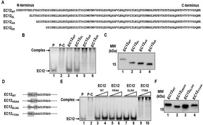
The N-terminal boundary in EC12 for binding of Cry1Ab. A series of deletion mutants at the N-terminal end of EC12 was constructed. Mutant proteins were purified by nickel affinity chromatography. Competition binding of the mutant peptides was assessed using fluorescently labeled EC12 probe and purified Cry1Ab toxin. (A) Sequences for EC12wt and the N-terminal deletional mutant peptides. (B) Samples from the competitive binding assay resolved on a native gel. Shown is a representative fluorescent image of a native gel displaying competitive binding of the EC12 mutants to Cry1Ab toxin. The Cry1Ab-EC12 complex and unbound EC12 probe are indicated by arrows on the left. P: 200 nM EC12 probe alone (lane 1); P-C: 200 nM EC12 probe plus 500 nM Cry1Ab; other labels at top of the image show the non-fluorescent 500 nM EC12wt (lane 3) and the N-terminal truncated mutant peptides (lanes 4–6) at the same molar concentration incubated with Cry1Ab prior to EC12 probe addition. (C) Western blot analysis of the peptide competitors using anti-6xHis antibody demonstrates expression of the peptides. (D) The mutants of EC12 in which amino acid residues were substituted by alanine are presented. Dashed lines represent unchanged EC12 amino acid sequences. All alanine substitution mutants were expressed and purified by nickel affinity chromatography. Assays were conducted to test the mutants’ ability to compete with the EC12 probe for CrylAb binding. (E) Fluorescent gel displays competitive binding of the mutants listed in D. Fluorescent Cry1Ab-EC12 complex and unbound EC12 bands are indicated by arrows on the left side of the gel. P: 200 nM EC12 probe only (lane 1); P-C: 200 nM EC12 probe plus 500 nM CrylAb (lane 2); non-fluorescent EC12wT (lanes 3 and 4: 400 nM and 800 nM, respectively), EC12VR/AA (lanes 5 and 6: 400 nM and 800 nM, respectively), EC12el/AA (lanes 7 and 8: 400 nM and 800 nM, respectively) and EC12YT/aa (lanes 9 and 10: 400 nM and 800 nM, respectively) were incubated with 500 nM CrylAb prior to the addition of the EC12 probe. (F) Western blot analysis using anti-6xHis antibody against EC12wt (lane 1) and the alanine substitution mutants (lanes 2–4) show expression of these peptides.

