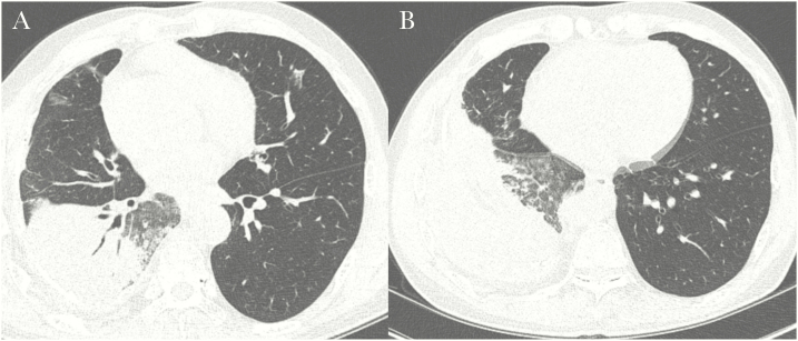
Figure 3.
A missed case of active pulmonary tuberculosis in a 58-year-old man with underlying liver cirrhosis and hepatocellular carcinoma. A, High-resolution computed tomography (CT) scan showing lobar consolidation with air-bronchogram and surrounding ground-glass opacities. Several patchy ground-glass opacities are evident in the right middle lobe and left upper lobe. B, Scan showing several ill-defined nodules in the right lower lobe. The CT findings were interpreted as nonspecific pneumonia, and the possibility of tuberculosis was neglected due to the lower lobe location and the main findings showing a consolidation with an air-bronchogram rather than discrete centrilobular nodules.

