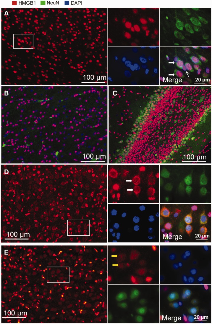
FIGURE 1.
Immunofluorescent staining of HMGB1 in sham-operated fetal sheep brain. (A) HMGB1 staining is positive in the cell nuclei in cerebral cortex. Double staining for HMGB1 and NeuN ( A , insert) shows that HMGB1 localizes to nuclei in both neuronal (wide arrows) and non-neuronal cells (narrow arrow). (B, C) Nuclear localization of HMGB1 was also observed in white matter (B) and hippocampus (C) . (D) Cells with cytoplasmic staining for HMGB1 were detected in the cerebral cortex of the sham-operated fetal sheep brain (insert). Double staining for HMGB1 and NeuN indicates neurons with cytoplasmic staining for HMGB1 (white arrows). (E) Neuronal cells (yellow arrows) with cytoplasmic staining for HMGB1 were also detected in normal fetal brains that had been perfused with formalin before brain collection (red, HMGB1; green, neurons/NeuN; blue, nucleus/DAPI).

