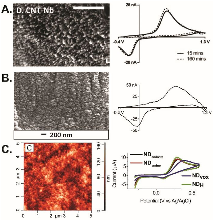
Fig. 2.
Carbon nanomaterial electrodes. (A) SEM image and FSCV of 1 μM dopamine at CNTs grown on Nb wire after 15 min (solid) and 160 min (dashed) of equilibration. Scale bar: 500 nm. Adapted with permission from Ref.6. Copyright 2016 American Chemical Society. (B) SEM image and FSCV of 1 μM dopamine at CNSs grown on Nb wire. Reproduced from Ref.28 with permission from The Royal Society of Chemistry. (C) AFM image of ND-coated taC electrodes and CVs of 1 mM dopamine at different types of nanodiamond. Reprinted from Ref.37 with permission from Elsevier.

