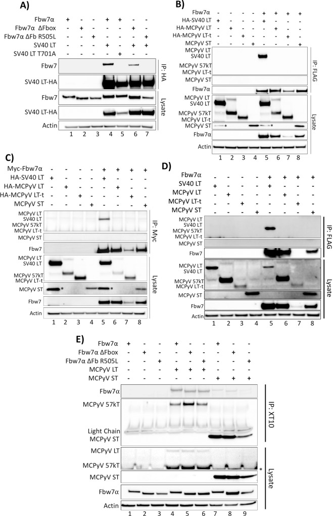
Fig 1. SV40 LT, but not the MCPyV T antigens, co-immunoprecipitate with Fbw7α.
(A) The SV40 LT antigen was pulled down with an anti-HA antibody from whole cell lysates of 293A cells expressing individual or combinations of HA-SV40 LT or the T701A mutant (5μg), wild-type FLAG-Fbw7 (4.5μg), or FLAG-Fbw7 ΔFbox/R505L mutants (3μg). Detection of co-immunoprecipitated Fbw7 was performed by immunoprecipitating with anti-HA, followed by immunoblotting with anti-FLAG. (B) The reciprocal IP to Fig 1A was performed with HA-SV40 LT, the MCPyV T antigens (HA-LT (5μg), HA-LT-t (10.5μg), and untagged ST (1μg)) and Fbw7, in which Fbw7 was pulled down (FLAG) and immunoblotted for interacting T antigens (anti-HA/2T2 (common-T antibody)). (C) An identical co-immunoprecipitation as Fig 1B was performed, except Fbw7 with an N-terminal Myc tag was pulled down from cellular lysates using a Myc tag specific antibody (9E10). (D) An identical co-immunoprecipitation as Fig 1B was performed with untagged SV40 and MCPyV T antigens. SV40 and MCPyV T antigens were detected by XT10 immunoblotting. (E) A co-immunoprecipitation between MCPyV T antigens (LT and ST) and Fbw7 was also performed through pull-down of the T antigens (XT10—common-T antibody) and detection of co-immunoprecipitated Fbw7 (anti-FLAG). Asterisks (*) denote non-specific bands.

