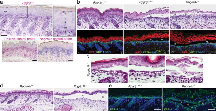
Fig 1. Intraepidermal blistering in Rpgrip1l-/- skin.
(a) In situ hybridization of Rpgrip1l in the dorsal skin of E18.5 wild-type mouse. Rpgrip1l (pink dot) is expressed in the epidermis (Epi), dermis (Der), and hair follicles (HF). Dotted line represents basement membrane. B, basal keratinocyte; SB, suprabasal keratinocyte; S, spinous keratinocyte; G, granular keratinocyte. Positive and negative control probes detect the mouse POLR2A or bacterial dapB gene, respectively. (b) Hematoxylin and eosin (H&E) staining and immunofluorescence labeling of KRT14 (green) and KRT1 (red) in dorsal skins of E18.5 control (Rpgrip1+/+) and homozygous mutants (Rpgrip1l-/-). Nuclei were labeled with DAPI (blue). Asterisks indicate intraepidermal blisters. (c) High power H&E images to demonstrate details of the blistering region. B, basal keratinocyte; SB, suprabasal keratinocyte; S, spinous keratinocyte. (d and e) H&E staining (d) and immunofluorescence labeling of KRT14 (green, e) of organotypic skin explants from E18.5 control (Rpgrip1l+/+, n = 8) and homozygous (Rpgrip1l–/–, n = 7) embryos at day 2. Nuclei were labeled with DAPI (blue). Asterisks indicate intraepidermal blisters. Scale bar, 25 μm in (a), 100 μm in (b), 10 μm in (c), 50 μm in (d, e).

