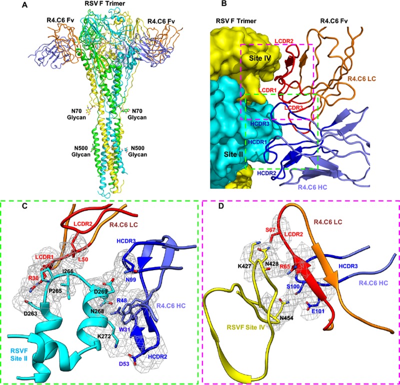
Fig 3. Structure of post-fusion RSV F710 in complex with R4.C6.

(A) Ribbon representation of the model of R4.C6 Fv bound to RSV F glycoprotein trimer in post-fusion conformation in side view. Each protomer of RSV F has a different color (cyan, yellow, and green). Only two of the three R4.C6 Fv molecules (light chain in orange color and heavy chain in blue color) are shown for clarity. The N-acetyl-D-glucosamine moieties attached to N70 and N500 are shown. (B) The interface between RSV F and R4.C6 heavy chain (HC, blue color) and light chain (LC, orange color). CDRs are labeled for R4.C6 Fv (HC, dark blue color; LC, red color). Antigenic site II and IV are also labeled. Two regions to be zoomed in (C) and (D) are highlighted by dashed squares. (C) Detailed interactions between R4.C6 and RSV F antigenic site II. (D) Detailed interactions between R4.C6 and RSV F antigenic site IV. RSV F site II is colored as cyan and site IV is colored as yellow. Cryo-EM maps around the residues in close proximity at the RSV F-R4.C6 interface are shown in grey mesh. The same color scheme is used in (B~D).
